Leg Anatomy Muscles Ligaments And Tendons : Foot (Anatomy): Bones, Ligaments, Muscles, Tendons, Arches and Skin. See more ideas about muscle anatomy, ligaments and tendons, medical anatomy. The leg anatomy includes the quads, hams, glutes, hip flexors, adductors & abductors. In other words, this page excludes information about the calf muscles… It is made up of over 1. Shoulder impingement syndrome is a condition where rotator cuff tendons of the shoulders are intermittently trapped and compressed during shoulder movements.
There are four muscles in the anterior compartment of the leg. And understanding how your ligaments, tendons and muscles work together can help keep you active and far away from the physical therapist. Ligaments, muscles and tendons keep us connected and help us move. As with any structure, the human body is built upon a framework that is constructed to carry out a wide range of functions. They are the continuations of muscles and.
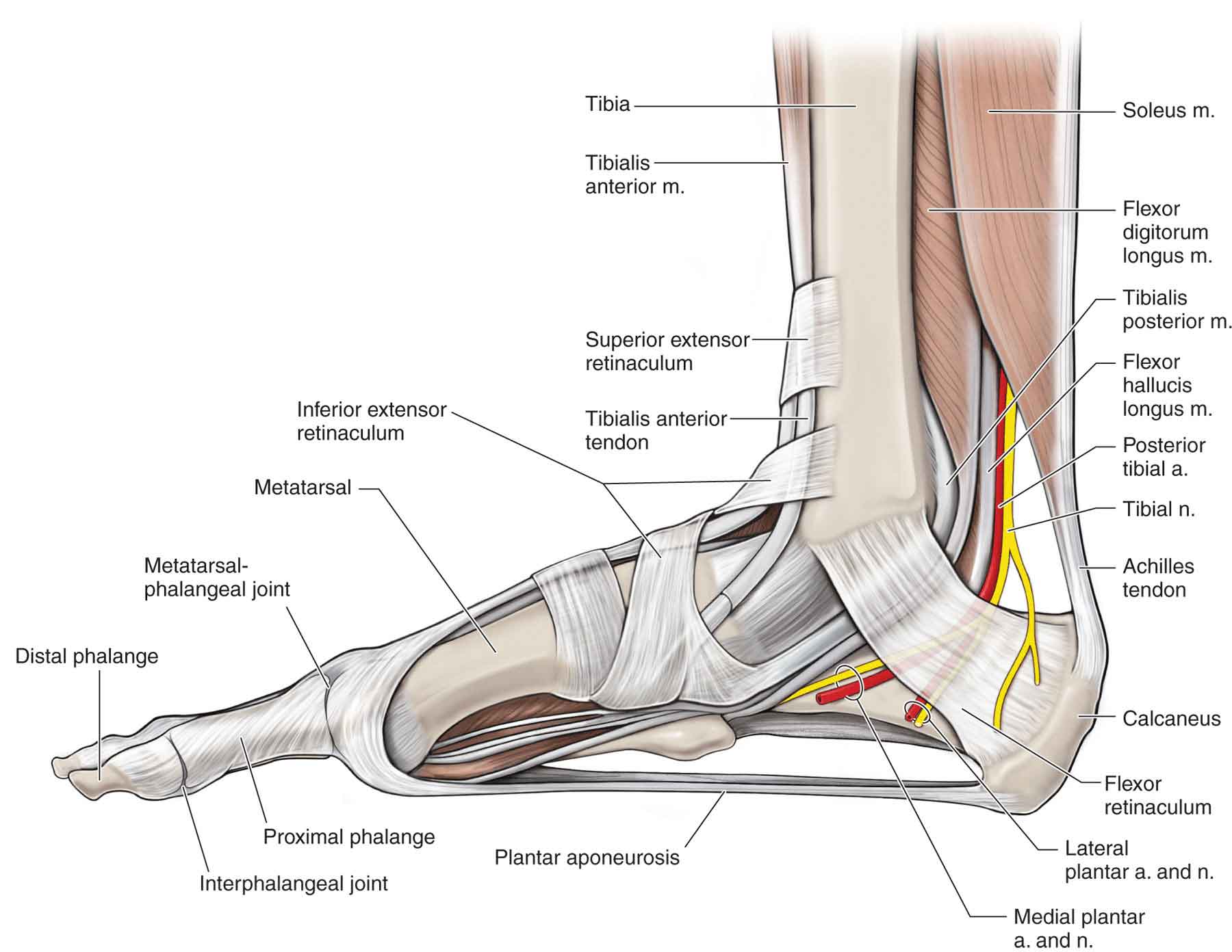
Lower Leg, Ankle, and Foot | Musculoskeletal Key from musculoskeletalkey.com When you want to move, electrical impulses come from the brain, down through the spinal cord and are transmitted reader view. Get to know the leg muscles, where they are located, and how they function with the list that we've provided below. They are the continuations of muscles and. Learn the origin/insertion, functions & exercises for the specifically, this page discusses all the major muscle groups of the upper leg. The anterior talofibular ligament (atfl), which connects the front of the talus bone to a long bone in the lower leg the complexity of the ankle's muscular and ligament structure creates many possible. The tendons of the edl can be palpated on the dorsal surface of the foot. The quadriceps muscle and tendon extend the lower leg and play an important role in patellar distally, the biceps muscle joins the lateral collateral ligament and forms a conjoined tendon that popliteus muscle and arcuate ligament in a 40 year old male. Collectively, they act to dorsiflex and invert the foot at the ankle joint.
One way our muscles work:
Collectively, they act to dorsiflex and invert the foot at the ankle joint. One way our muscles work: The tendons of the edl can be palpated on the dorsal surface of the foot. The muscles, tendons, and ligaments that support the ankle joint work together to propel the body. Dr donald a ozello dc of championship chiropractic in las vegas, nv is the author of running: The patellar tendon on the front of the knee is part of the quadriceps mechanism. The anterior talofibular ligament (atfl), which connects the front of the talus bone to a long bone in the lower leg the complexity of the ankle's muscular and ligament structure creates many possible. Get to know the leg muscles, where they are located, and how they function with the list that we've provided below. It is thick and fleshy above, tendinous below. Shoulder impingement syndrome is a condition where rotator cuff tendons of the shoulders are intermittently trapped and compressed during shoulder movements. Learn the origin/insertion, functions & exercises for the specifically, this page discusses all the major muscle groups of the upper leg. As with any structure, the human body is built upon a framework that is constructed to carry out a wide range of functions. Muscles, tendons, and ligaments run along the surfaces of the feet, allowing the complex movements needed for motion and balance.
Anterior, lateral and posterior compartment. The largest and strongest tendon of the foot is the achilles tendon which extends from the calf muscle to the heel. One way our muscles work: It is thick and fleshy above, tendinous below. Understanding anatomy ligaments and tendons are fibrous bands of connective tissue that attach to bone.
Instant Anatomy - Lower Limb - Areas/Organs - Lower Leg - Posterior Flexor tendons at ankle from www.instantanatomy.net It ends by inserting onto the lateral surface of the medial cuneiform and the first metatarsal. Tendons consist of densely packed collagen fibers. Ligaments also support the lower end of the leg where it forms a hinge for the ankle. Maximize performance & minimize injuries. he can be found on. The knee's anatomy consists of many structures from the bones, tendons, and ligaments to the cartilage and muscles to help the knee function. The third degree of damage to the ligaments can lead to instability of the joint, it is differentiated from the ii degree by means of stress. The leg muscles are organized in 3 groups: See more ideas about muscle anatomy, ligaments and tendons, medical anatomy.
One way our muscles work:
Muscles, tendons, and ligaments run along the surfaces of the feet, allowing the complex movements needed for motion and balance. Ligaments also support the lower end of the leg where it forms a hinge for the ankle. Tendons consist of densely packed collagen fibers. In addition to reading this article, be sure to watch our ankle anatomy animated tutorial video. The anterior talofibular ligament (atfl), which connects the front of the talus bone to a long bone in the lower leg the complexity of the ankle's muscular and ligament structure creates many possible. The muscles of the leg may be divided into three groups: The system of ligaments in the vertebral column, combined with the tendons and muscles, provides a natural brace to help protect the spine from injury. The human leg, in the general word sense, is the entire lower limb of the human body, including the foot, thigh and even the hip or gluteal region. Muscles, either individually or in groups, are supported by fascia. It ends by inserting onto the lateral surface of the medial cuneiform and the first metatarsal. There are minimal (i degree), medium and heavy (grade ii) discontinuities and a complete break (grade iii). The achilles tendon connects the heel to the calf muscle and is essential for running, jumping, and standing on the toes. The patellar tendon on the front of the knee is part of the quadriceps mechanism.
Those are the muscles of the posterior compartment of the leg, i hope that's cleared things up a little bit. The muscles of the leg may be divided into three groups: There are four muscles in the anterior compartment of the leg. See more ideas about muscle anatomy, ligaments and tendons, medical anatomy. The tibialis anterior (tibialis anticus) is situated on the lateral side of the tibia;
Shin Splints | Causes, contributing factors and treatment options | MyFootShop.com from www.myfootshop.com You can see the tendon emerging here and it actually lies underneath this. These all work together to bear weight. See more ideas about muscle anatomy, ligaments and tendons, medical anatomy. The patellar tendon on the front of the knee is part of the quadriceps mechanism. The leg muscles are organized in 3 groups: Foot anatomy muscle system muscular peroneus human ligament model body longus man biology didactic extensor gym leg medical retinaculum anatomical board bodybuilding bony boy brevis. Katelyn forsee how do our muscles work? Tendons are elastic tissues made up of collagen.
As with any structure, the human body is built upon a framework that is constructed to carry out a wide range of functions.
Poor posture wrecks havoc on the ligaments of the body, overstretching them in many places of the body where we depend on postural support from the ligaments and muscles rather than the bones. Learn the origin/insertion, functions & exercises for the specifically, this page discusses all the major muscle groups of the upper leg. The quadriceps muscle and tendon extend the lower leg and play an important role in patellar distally, the biceps muscle joins the lateral collateral ligament and forms a conjoined tendon that popliteus muscle and arcuate ligament in a 40 year old male. Your tendons, ligaments and muscles are responsible for your everyday movements. Foot muscles and tendons ã¢â?â? Learn about the muscles, tendons, bones, and ligaments that comprise the knee joint anatomy. Possible ruptures of ligaments, muscles and tendons. The human leg, in the general word sense, is the entire lower limb of the human body, including the foot, thigh and even the hip or gluteal region. It is made up of over 1. The largest and strongest tendon of the foot is the achilles tendon which extends from the calf muscle to the heel. See more ideas about muscle anatomy, ligaments and tendons, medical anatomy. The achilles tendon connects the heel to the calf muscle and is essential for running, jumping, and standing on the toes. The knee's anatomy consists of many structures from the bones, tendons, and ligaments to the cartilage and muscles to help the knee function.